


BibTeX | RIS | EndNote | Medlars | ProCite | Reference Manager | RefWorks
Send citation to:
URL: http://jsurgery.bums.ac.ir/article-1-162-en.html


Surgical removal of a foreign body from esophagus: in case of endoscopic removal failure
Malihe Zanguoie *
Assistant Professor, Department of Anesthesiology, faculty of Medicine, Birjand University of Medical Sciences, Birjand, Iran
Received: July 15, 2018 Revised: August 25, 2018 Accepted: October 17, 2018
|
Abstract Foreign body ingestion is remarkable in adults although the incidence of this issue is more common in children. The complication level of foreign body ingestion depends on the site and type of foreign body. Moreover, partial denture with sharp metal hooks, metal springs, and sharp bone pieces are among the most difficult and hazardous foreign bodies result in fistula formation or esophagus puncture. The reported case in this study was a man with a sharp steel wire in his throat. Rigid and flexible endoscopy are the interventions for the removal of foreign bodies; however, surgery is regarded as a safer and more effective treatment for difficult and hazardous cases. Key Words: Esophagus, Foreign Bodies, Impacted foreign body |
Introduction
Foreign body ingestion is common in infants aged six months to three years (1). The most common foreign bodies ingested by infants are coins, toys, jewelries, magnets, and batteries (2). On the other hand, considering adults, the common foreign ingested bodies are fish bone and other bone fragments, dentures and metallic wires (3).
Approximately 70-80 % of the swallowed foreign bodies will generally pass without need for intervention (4) and the flat foreign bodies tend to pass spontaneously as well. However, sharp foreign bodies stuck in esophagus may get it impacted or perforated in case they are not removed immediately. Moreover, sharp objects even pass uneventfully once they cross the esophagus (5).
Foreign body ingestion is more common in adults with mental disorders. Rigid and flexible endoscopy are regarded as the interventions for the removal of foreign bodies and they are applied for 20% of cases (6). On the one hand, the rigid endoscopy intervention is easy and safe; however, it requires general anesthesia.
On the other hand, flexible endoscopes have been used recently for removal of foreign bodies under local anesthesia with identical achievement. Moreover, the need for surgical intervention is realized in 16-12% of the patients. It is declared that postponement in presentation increases the risk of serious complications, such as impaction, and perforation (5). With this background in mind, this case report study evaluated a patient referring to hospital with the ingestion of a steel wire while eating food.
Cases
The case study was a 49-year-old man who swallowed a steel wire with his food and suffered from impaction and odynophagia. According to the endoscopic findings, a needle-shaped small bone object with inflammation was observed to be stuck in the upper part of esophagus. Subsequently, a part of the foreign body was taken out; however, a feeling of impaction and odynophagia was likely to


Figure1: The removed steel wire
All subsites of larynx were checked, and no complications except for mild inflammation in arytenoid cartilage were left. Furthermore, there was no evidence of laceration of mucous layer or carotid sheet or esophagus. Subsequently, with a separate approach in 3 cm upper part of supra notch, the flap was elevated in sub platisma position and sternocleidomastoid muscle was pulled over and behind of the carotid was explored. The surgeon chose the approach of medial behind the carotid, and the pre-vertebral fascia was exposed, then the fascia tissue and cervical esophagus were explored.
Behind the Carotid and esophagus, was traumatic and the foreign body with position of lateral to esophagus' wall was disclosed. The irrigation was performed and the drain was placed and NGT was fixed.
Discussion
Pediatric populations are more vulnerable to foreign body ingestion; however, this incidence is also remarkable in adults. The complication level of foreign body ingestion depends on the site and type of foreign body. Moreover, the most hazardous foreign bodies which result in fistula formation or esophagus puncture included sharp metal hooks, metal springs, and sharp bone pieces.
The reported case in this study was a man who swallowed a sharp steel wire while he was eating food. The results of the recent studies showed that the foreign objects should be removed within 24 h after the incidence. In addition, foreign body ingestion leads to 3.2% complication within 24 h after ingestion following by 23.5% increase after 48 h (7).
In the current study the patient underwent treatment within 24 h after the incidence. The neck and chest X-ray is considered as the non-endoscopic detection methods shown in figure 2 (8); moreover, rigid and flexible endoscope are regarded as interventions for removal of foreign bodies. The success rate with the use of rigid esophagoscopy is estimated at 94% to 100 % (9), on the other hand, this range is from 76% to 98.5 % for flexible esophagoscopy (3).
However, flexible esophagoscopy is more suitable for patients with the high risk of anesthetic. According to the results, the foreign bodies that are less than 7 mm, seldom cause trauma or severe damage (10). These findings are not compatible with the 20 mm ingested object of the current case led to surgery operation.
If endoscopic removal fails or the ingested objects are sharp ones to pierce the lumen, esophagotomy with surgery should be considered (9). Both symptoms were existed in the present case, therefore, the patient had to undergo surgery operation after the failure of endoscopic technique. The foreign body was removed successfully after the surgery.
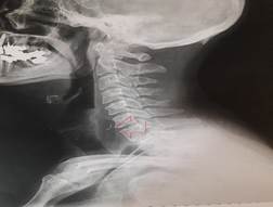
Figure2: The neck and chest X-ray of the foreign body
Conclusions
Steel wire impaction is a kind of difficult and dangerous foreign body. The most common way to encounter foreign objects is endoscopic removal; however, in this case it was removed by surgery operation due to the object sharpness. The surgery operation is recommended for sharp objects which penetrated into the esophagus and it is considered as a safer and more effective treatment.
Conflict of Interest
None Declared.
References
1. Uyemura MC. Foreign body ingestion in children. Am Fam Phys. 2005; 72(2):287-91.
2. Kramer RE, Lerner DG, Lin T, Manfredi M, Shah M, Stephen TC, et al. Management of ingested foreign bodies in children: a clinical report of the NASPGHAN Endoscopy Committee. J Pediatr Gastroenterol Nutr. 2015; 60(4):562-74. DOI: 10.1097/MPG.00000000
00000729
3. Mondal PJ, Saha S, Ghosh A, Sengupta M. Removal
of foreign bodies from esophagus with flexible endoscope-a case report. Ind J Otolaryngol Head Neck Surg. 2014; 66(1):78-80. PMID: 24533363 DOI: 10.1007/s12070-011-0320-9
4. Shivakumar AM, Naik AS, Prashanth KB, Hongal GF, Chaturvedy G. Foreign bodies in upper digestive tract. Indian J Otolaryngol Head Neck Surg.
2006; 58(1):63-8. PMID: 23120240 DOI: 10.1007/
BF02907744
5. Pillai SA, Sivasankar A, Selvaraj T. Foreign bodies in the oesophagus-surgery for failed endoscopic retrieval. Int Surg J. 2016; 3(3):1426-30. DOI: http://dx.doi.org/10.18203/2349-2902.isj20162722
6. Ambe P, Weber SA, Schauer M, Knoefel WT. Swallowed foreign bodies in adults. Deutsch Ärztebl Int. 2012; 109(50):869-75. PMID: 23293675 DOI: 10.3238/arztebl.2012.0869
7. Yan XE, Zhou LY, Lin SR, Wang Y, Wang YC. Therapeutic effect of esophageal foreign body extraction management: flexible versus rigid endoscopy in 216 adults of Beijing. Med Sci. 2014; 20:2054-60. PMID: 25349897 DOI: 10.12659/
MSM.889758
8. Dereci S, Koca T, Serdaroğlu F, Akçam M. Foreign body ingestion in children. Turk Pediatr Ars. 2015; 50(4):234-40. PMID: 26884693 DOI: 10.5152/
TurkPediatriArs.2015.3164
9. Shreshtha D, Sikka K, Singh CA, Thakar A. Foreign body esophagus: when endoscopic removal fails. Indian J Otolaryngol Head Neck Surg. 2013; 65(4):380-2. PMID: 24427604 DOI: 10.1007/
s12070-013-0662-6
10. Okhakhu AL, Ogisi FO. An unusual foreign body in human oesophagus–case report. Benin J Postgrad Med. 2007; 9(1):41-3. DOI: 10.4314/bjpm.v9i1.
47371
Received: 2018/07/15 | Accepted: 2018/10/17 | Published: 2018/11/14
| Rights and permissions | |
 |
This work is licensed under a Creative Commons Attribution-NonCommercial 4.0 International License. |

Attribution-NoneCommercial CC BY-NC
