


BibTeX | RIS | EndNote | Medlars | ProCite | Reference Manager | RefWorks
Send citation to:
URL: http://jsurgery.bums.ac.ir/article-1-189-en.html


Non-surgical and surgical management of four maxillary teeth with irreversible pulpitis secondary to a large inflammatory dentigerous cyst: A case report
Soheila Darmiani1*![]()
1Assistant professor in Endodontics, Department of Endodontics, Faculty of Dentistry, Birjand University of Medical Sciences, Birjand, Iran
Received: May 21, 2019 Revised: July 09, 2019 Accepted: August 06, 2019
|
Abstract Dentigerous cyst which is the most common developmental cyst of the jaw forms around the crown of an unerupted or supernumerary tooth. This case report demonstrates the treatment of a large inflammatory dentigerous cyst caused by an abnormal supernumerary tooth and the subsequent irreversible pulpitis affecting the four maxillary teeth. Key words: Dentigerous cyst, Maxilla, Pulpitis, Root canal therapy, Surgery, Tooth |
Introduction
Dentigerous cyst (DC) which is the second most common type of odontogenic cyst and the most common developmental cyst of the jaw (1) forms around the crown of an unerupted or supernumerary tooth. It emerges with fluid accumulation in the layers of reduced enamel epithelium or between the epithelium and the crown of the unerupted tooth (2). The occurrence of DC with supernumerary teeth was first described by Pitts in 1924(3).The highest incidence of DC occurs during the second and third decades of life with a greater incidence detected in males (4). Patients usually have no complaint of pain or discomfort and the clinical examination demonstrates a hard swelling which sometimes leads to facial asymmetry (5). Radiographically, DC presents as a well-defined unilocular radiolucency with corticated margins in association with the crown of an unerupted tooth. The epicenter of a DC is most commonly the mandibular, maxillary third molar, or the maxillary canine. Extension into the maxillary sinus or the orbital floor may be detected in maxillary DCs involving the canine region. In addition, resorption of roots of adjacent erupted teeth may occasionally be detected (6). An important diagnostic point is that this cyst attaches to the cementoenamel junction (CEJ).The final diagnosis is only feasible following histological examination. Histologically, the DC consisted of a fibrous connective tissue wall and is lined by a stratified squamous epithelium (4). In an uninflamed DC, the epithelial lining is non-keratinized and tends to be approximately four to six cell-layer thick, although epithelial hyperplasia may be noted in cases of secondary inflammation. DC has a good prognosis (3,6) and
is treated by enucleation or marsupialization/ decompression methods.
This case report recounts the successful treatment of a large inflammatory DC caused by
an abnormal supernumerary tooth and the subsequent irreversible pulpitis affecting the four maxillary teeth.
Case
A 33-year-old male presented to the Diagnosis

Figure1: Preoperative periapical radiograph of teeth # 10, 11, 12 and 13
Department of the Shahid Beheshti University of Medical Sciences, Tehran, Iran with a complained of a painless, slow-growing asymptomatic facial swelling that had started 9 months ago. The swelling was initially of a small size and gradually increased to its present size. There was no associated history of trauma, syndromes or systemic diseases. The medical history was noncontributory and his body temperature was measured at 37.5ºC. Extraoral examination evidenced swelling in the left anterior region of the maxilla. In addition, palpation revealed that swelling was non- tender and firm inconsistency. The teeth #10, 11, 12, and 13 responded normally to both percussion and palpation tests. Moreover, the periodontal probing was within the
normal range (<3mm). Furthermore, the teeth demonstrated an exacerbated response to electrical and thermal tests, as compared to control teeth # 4, 5, 7. The results of the tests confirmed the diagnosis of irreversible pulpitis. A periapical radiograph (Figure 1( demonstrated a unilocular well-defined radiolucent lesion which involved periapical regions of teeth # 10, 11, 12, and 13. The radiolucent lesion circumscribed the cement-enamel junction (CEJ) of the impacted supernumerary tooth crown. It is worthy to note that patient's posterior-anterior (PA) cephalometric radiograph was also available (Figure 2).
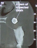
Fine-needle aspiration (FNA) demonstrated blood-tinged cystic fluid with cholesterol crystals revealed by examination. The patient underwent cone-beam computed tomography (CBCT) for
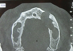
more detailed examination of the finding and investigation of the proximity or any involvement of lesion with nasal floor and the maxillary sinus due to lesion extension (Figures 3 A-D). The CBCT evidenced the erosion and buccal cortex perforation, cortical thinning of the floor of nasal cavity and maxillary sinus. Provisional diagnosis of
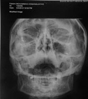
Figure 2: Radiolucent mass identified in the PA cephalometric projection
dentigerous cyst was made based on history and clinical examination. The differential diagnosis included dentigerous cyst, odontogenic keratocyst (OKC), calcifying odontogenic cyst (COC), adenomatoid odontogenic tumor (AOT), and ameloblastoma.
Root canal therapy was the treatment plan suggested to the patient; thereafter, he was provided with the necessary explanation and informed written consent was obtained from him.The patient received an infiltration injection
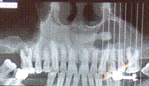
of 2% lidocaine with 1:80000 epinephrine (DarouPakhsh, Tehran, Iran). Thereafter, access cavity was prepared and canals orifices were identified. The working length (WL) was estimated using Root ZX apex locator (J. Morita USA, Inc., Irvine, CA, USA) and a radiograph was performed to confirm the WL (Figure 4). Mechanical preparation of the roots canals was carried out using a crown down technique by HERO 642 rotary files (Micromega, France) and motor controller device(X Smart, Dentsply, Maillefer, Ballaigues, Switzerland) in the following sequences: 20.02, 25 02,25.04, 30.02, 30.04. Moreover, Saline and 2.5% NaOCl were used for repeated irrigation. The roots canals were dried with paper points (Ariadent, Tehran, Iran) and obturated with gutta-percha (Ariadent, Tehran, Iran) and AH-26 sealer (Dentsply, DeTrey, Konstanz, Germany) using cold lateral condensation technique (Figure 5) and the patient was referred for permanent restoration.
The patient was scheduled for surgery in the next visit which included surgical enucleation of lesion, along with the removal of the impacted supernumerary tooth, apicoectomy, and retrograde filling of involved teeth. Routine blood tests run prior to surgery revealed normal values. The surgery was performed in Surgery Department of Shahid Beheshti University of Medical Science,
(A) (B)

(C) (D)
Figures 3(A-d): Axial CBCT views demonstrating extent of lesion, perforation of the buccal cortical plate. Coronal CBCT view depicting displacement of the sinus membrane (suggests a sinus membrane perforation will be detected during surgery)
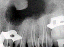
Figure 4: Working length determination Radiograph
Tehran, Iran under local anesthesia. Crevicular incision was conducted on labial region teeth #9-14. A full-thickness mucoperiosteal flap was reflected, the cyst was exposed and complete curettage was performed. The rupture of the nasal floor was immediately closed by resorbable suture (vicryl 5-0; Figure 6).The biopsy was conducted and sent for histological examination. The apical 3 mm of the roots were resected perpendicular to the long axis of the teeth with a diamond fissure bur (010, Tizkavan, Tehran, Iran) in high-speed dental handpiece under water spray. Three-millimeter deep root-end cavities were prepared with a diamond-coated retro-tip attached to an ultrasonic unit (Varios 970, NSK, Japan) and retrograde filling was performed with CEM cement
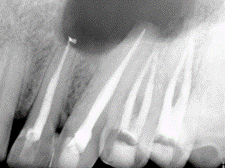
Figure 5: Final radiography after root canal obturation
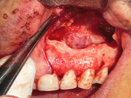
Figure 6: Clinical image after surgical access removal of lesion and root-end resection demonstrating sinus perforation
(A) (B)
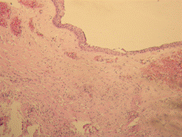
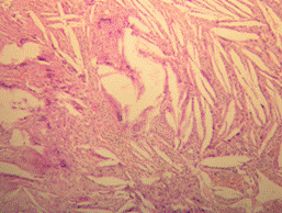
Figure 7: A-B.Light microscopic section of the biopsy specimen (×400), the lesion was diagnosed as an inflammatory dentigerous cyst.
(BioniqueDent, Tehran, Iran) which was prepared according to manufacturer's instructions. CEM cement mixture was delivered into the root end cavities and gently packed with appropriate pluggers and paper points. Flap closure was accomplished with 3-0 silk sutures. A TID prescription of 500 mg Amoxicillin capsules for 7 days and on time consumption of 400mg ibuprofen supplemented the treatment and the patient was discharged after receiving necessary instructions. The patient was recalled 4 days
after surgery for sutures removal and he was rechecked after 10 days. The clinical signs/symptoms had resolved and the histological examination revealed a cyst wall composed of fibrous tissue and lined by non-keratinized stratified squamous epithelium with hyperplasia in some area (Figure 7). An inflammatory DC was considered the final diagnosis after correlating the history, clinical andradiographical features, along with histopathology. Currently, the patient is asymptomatic and no recurrence and favorable osseous formation were observed during the 2 years follow-up period (Figure 8).
Discussion
The term “cyst”, which is derived from the Greek word “Kystis”, meaning, “sac or bladder”, is defined as a pathological cavity usually lined by epithelium (4).
A DC surrounds the crown of an unerupted tooth, expands the follicle, and notably attaches to the CEJ of the unerupted tooth (6). The cyst commonly influences people within the age group of 10-30 years with males being more affected with an incidence rate of 1.6:1. In addition, DC usually occurs throughout the first four decades of life due to supernumerary tooth. Despite the
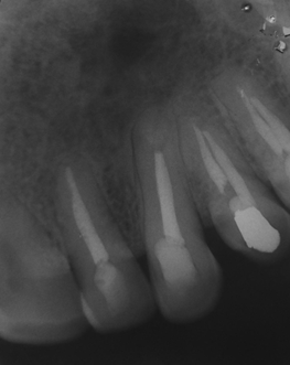
Figure 8: The 2-year follow-up radiography
solitary nature of DCs, multiple cysts might be detected with such syndromes as Gardner’s syndrome, mucopolysaccharidosis, Maroteaux-Lamy syndrome, and basal cell nevus syndrome. Patients commonly present with unerupted teeth or asymptomatic slow-growing swelling. In this case, the patient was a 36-year-old male who presented with an asymptomatic solitary swelling in the left anterior region of the maxilla.
Radiographic examination of DC demonstrated a unilocular radiolucent lesion associated with the crown of an unerupted tooth and well-defined sclerotic margins. However, CBCT is required in cases of the extensive lesion (7). CBCT provides information on origin, size, cortical plates, and relationship of the lesion with adjacent anatomical structures (8). CBCT also was obtained in this case on account of the size of the lesion.
Although DCs are benign in nature, they can get quite destructive if they go undetected due to their expansive nature, as demonstrated in this case. The neighboring teeth can possibly develop endodontic disease in the form of reversible and irreversible pulpitis. Apart from the expansion of DC, loss of vitality can result from disruption of blood flow at the apex of the affected tooth (5,6).This case indicated that the teeth with irreversible pulpitis secondary to pressure from a DC can be successfully managed.
Surgical endodontic literature supports a direct cause and effect relationship between the hermetic sealing of the apical area by root-end filling materials and successful outcomes. The ideal root-end filling material seals the contents of the root canal system within the canal and prevents the egress of any bacteria, bacterial byproducts, or toxic material into the surrounding periradicular tissues. The material should be nonresorbable, biocompatible, and dimensionally stable over time. It should be able to induce regeneration of the PDL complex, specifically cementogenesis over the root-end filling itself. Many materials have been used as root-end fillings, including guttapercha, polycarboxylate cement, amalgam, zinc oxide eugenol(ZOE) cement (IRM and SuperEBA), glass ionomer cement, Diaket, composite resins (Retroplast), resin–glass ionomer hybrids (Geristore), MTA, and CEM cement(9,10). Sealing ability of CEM cement is comparable to those of MTA and since these two water-based endodontic biomaterials do not demonstrate shrinkage upon setting, they form hydroxyapatite crystals over their surfaces. The results of several studies conducted specifically on regenerative endodonticsand vital pulp therapy were indicative of favorable biocompatibility of CEM cement. Moreover, randomized controlled trials have suggested that cementogenic properties of CEM cement are comparable to those of MTA when used as a root-end filling material (9).
Conclusions
Based on the obtained results, the timely detection and treatment of impacted tooth are of paramount importance to impede the likely expansion of a DC. At the same time, if a DC diagnosis prolongs until substantial expansion, therapeutic measures for the treatment of affected teeth is crucial.
Acknowledgments
Our sincere appreciation and thanks go to Pathology and Surgery Department of Shahid Beheshti University of Medical Sciences. Moreover, we acknowledge the valuable contribution of all participants.
Conflict of Interest
The authors declare that no competing interests exist.
References
1. Huang G, Moore L, Logan RM, Gue S. Histological analysis of 41 dentigerous cysts in a paediatric population. J Oral Pathol Med. 2019; 48(1):74-8. PMID: 30175860 DOI: 10.1111/jop.12776
2. De França GM, Cavalcante IL, Barros CC, Junior LC, Germano AR, Queiroz LM, et al. Dentigerous cyst of inflammatory origin in a pediatric patient: a case report. Oral Surg Oral Med Oral Pathol Oral Radiol. 2018; 126(3):e112. DOI: 10.1016/j.oooo.2018.
02.396
3. Pitts AT. A Dentigerous cyst apparently associated with a supernumerary tooth. Proc R Soc Med. 1924; 17:9-10. PMID: 19983891
4. Buchbender M, Neukam FW, Lutz R, Schmitt CM. Treatment of enucleated odontogenic jaw cysts: a systematic review. Oral Surg Oral Med Oral Pathol Oral Radiol. 2018; 125(5):399-406. PMID: 29396318 DOI: 10.1016/j.oooo.2017.12.010
5. Marino MJ, Luong A, Yao WC, Citardi MJ. Management of odontogenic cysts by endonasal endoscopic techniques: a systematic review and case series. Am J Rhinol Allergy. 2018; 32(1):40-5. PMID: 29336289 DOI: 10.2500/ajra.2018.32.4492
6. Mello FW, Melo G, Kammer PV, Speight P, Rivero ER. Prevalence of odontogenic cysts and tumors associated with impacted third molars: a systematic review and meta-analysis. J Craniomaxillofac Surg. 2019; 47(6):996-1002. PMID: 31005378 DOI: 10.1016/j.jcms.2019.03.026
7. Gao L, Ren W, Li S, Zheng J, Xue L, Xu Y, et al. CBCT-based bone quality assessment in decompression of large odontogenic cystic lesions. Oral Radiol. 2018; 34(3):251-6. PMID: 30484038 DOI: 10.1007/
s11282-018-0320-5
8. Meng Y, Zhang YQ, Ye X, Zhao YN, Chen Y, Liu
DG. Imaging analysis of ameloblastoma, odontogenickeratocyst and dentigerous cyst in the maxilla using spiral CT and cone beam CT. Zhonghua kou Qiang Yi Xue Za Zhi. 2018; 53(10):659-64.
PMID: 30392221 DOI: 10.3760/cma.j.issn.1002-0098.2018.10.003
9. Francis T, Joshi SB, Pai AV, Sakkir N, Thaha KA. Comparison of the sealing ability of MTA-angelus, biodentine and CEM cement in the repair of large furcal perforations-a bacterial leakage study. J Clin Diagn Res. 2019; 13(1):32-5.
10. Torreira MG, Dos Santos AA, Cobos MR, Boquete IF, Abelleira AC. The osteoinductive potential of MTA (Mineral trioxide aggregate): a histologic study in rabbits. Eur J Anat. 2004; 8(3):101-5.
Received: 2019/05/21 | Accepted: 2019/08/6 | Published: 2020/02/17
| Rights and permissions | |
 |
This work is licensed under a Creative Commons Attribution-NonCommercial 4.0 International License. |

Attribution-NoneCommercial CC BY-NC
