

Volume 13, Issue 3 (9-2025)
J Surg Trauma 2025, 13(3): 120-123 |
Back to browse issues page


Download citation:
BibTeX | RIS | EndNote | Medlars | ProCite | Reference Manager | RefWorks
Send citation to:


BibTeX | RIS | EndNote | Medlars | ProCite | Reference Manager | RefWorks
Send citation to:
Shahraki M, Ebrahimgol S, Hatampour S, Amirpour Haradasht S. Unusual Presentation of Salivary Stone: A Case Report of a Large Submandibular Duct Calculus Presenting as a Sore Throat in a Middle-aged Male. J Surg Trauma 2025; 13 (3) :120-123
URL: http://jsurgery.bums.ac.ir/article-1-457-en.html
URL: http://jsurgery.bums.ac.ir/article-1-457-en.html


Department of Oral and Maxillofacial Surgery, School of Dentistry, Zahedan University of Medical Sciences, Zahedan, Iran
Full-Text [PDF 410 kb]
(704 Downloads)
| Abstract (HTML) (1531 Views)

Discussion
Salivary gland inflammation due to stones is common, especially in the submandibular gland. Determining the true prevalence of salivary stones can be challenging, as they are often asymptomatic. Factors contributing to stone formation include saliva flow issues and composition. Duct irregularities, inflammation, dehydration, and certain medications lead to flow issues, while calcium levels and a lack of crystallization inhibitors affect saliva composition. Bacterial infections can also promote stone formation by increasing saliva saturation with calcium phosphate due to pH changes. Factors, such as duct shape, calcium levels, gland position, mucus, smoking, trauma, infection, and inflammation, contribute to salivary stasis. The recurrence rate is about 20%. The stone formation mechanism is unclear but likely involves microcalcifications due to reduced secretion over time in ducts (1, 7, 8).
Salivary stones can lead to intermittent swelling, with the severity often depending on the degree of obstruction and the presence of infection. While symptoms often occur during meals, this was not the case with our patient. Chronic cases may exhibit fistulas or ulcers, which were absent in our case. Submandibular stones are generally singular, whereas sublingual stones tend to be multiple and scattered. If untreated, stones can grow larger and eventually lead to calcification. Obstruction can also trigger infections, with symptoms including pain, swelling during meals, and infection-related symptoms in the case of larger stones (1, 3, 7).
In the acute phase, treatment for sialoliths focuses on supportive care, such as pain relief, hydration, and antipyretics, along with antibiotics to address potential infections caused by salivary gland inflammation. Stimulating saliva flow and localized massage therapy are commonly employed, with treatment options ranging from minimally invasive to more radical interventions depending on the size and characteristics of the stone (5, 9).
Factors influencing treatment decisions include the duration of obstruction, tissue changes, and stone location. Non-invasive methods, such as massage and medications, are effective for stones located more distally, while deeper stones often require surgical intervention. For submandibular gland stones, lithotripsy ultrasound is a well-established treatment. Sialendoscopy and laser lithotripsy can also be used for stones of varying sizes. In the differential diagnosis of masses in the floor of the mouth, it is essential to consider conditions, such as ranula, cysts, tumors, and calcifications in the lymph nodes, ducts, and glands (1, 3, 5).
Conclusions
Salivary stones are a known cause of salivary gland dysfunction and can present with a variety of clinical manifestations, including pain, swelling, and infection. However, the presentation of a salivary stone as a sore throat is uncommon and can mimic other more common conditions, necessitating a high index of suspicion for accurate diagnosis. This case underscores the importance of considering salivary stones in the differential diagnosis of patients presenting with atypical symptoms in the head and neck region. Prompt recognition and appropriate management of salivary stones can lead to favorable outcomes and symptom resolution. Further studies and case reports are warranted to enhance our understanding of the diverse presentations and optimal management strategies for this condition.
Conflict of Interest
The authors declare no conflicts of interest.
Full-Text: (351 Views)
Abstract
Salivary stones, or sialoliths, are calcified masses that can obstruct the ducts of the salivary glands, resulting in various clinical manifestations. This case report details the rare occurrence of a large submandibular duct calculus in a 42-year-old male who initially presented with sore throat symptoms. The patient’s clinical history, physical examination, imaging findings, and treatment approach are thoroughly outlined. This case underscores the necessity of including salivary stones in the differential diagnosis when managing patients with unusual symptoms in the head and neck region, particularly in those with atypical sore throat presentations.
Key words: Salivary Duct Calculi, Salivary Gland Calculi, Submandibular Gland
Introduction
Sialolithiasis, also known as salivary gland stones, is an organic calcified substance that forms in the paranchyma and ducts of both major and minor salivary glands; however, it usually involves a single gland. Mechanical factors that impede saliva flow, along with the physicochemical properties of gland secretions, contribute to the formation of a central core for calcification and subsequent deposition of calcium salts and phosphates. The condition is most common in adult males aged 30 to 60, with a lower prevalence in children. The precise etiology remains unclear, though potential contributing factors include inflammation, infection, anatomical irregularities in the salivary duct system, and smoking (1, 2).
Sialolithiasis predominantly affects the submandibular gland and its duct due to its complex structure, while less common occurrences are seen in the parotid gland and the sublingual or minor salivary glands. The clinical importance lies in recognizing these patterns, particularly when faced with atypical presentations that may challenge the initial diagnosis (3). It should be noted that the mucoid nature of submandibular gland secretions with high levels of calcium and phosphate makes sialolithiasis more common in this gland. Secretions from submandibular and parotid glands are nerve-dependent; accordingly, when nerve stimulation is absent, the risk of sialolith formation increases. The size of these calcified stones varies greatly (4).
Multiple stones in more than one salivary gland are scarce (about 3%). They can have various shapes, such as spherical, oval, or irregular. Symptoms can also vary, and some patients may be asymptomatic while others may experience severe pain, especially when eating. Saliva production may decrease in some patients. Swelling and inflammation around the duct entrance, oral mucosa floor, and skin near the salivary gland area may occur (5).
The severity of symptoms is influenced by the location and extent of saliva flow obstruction. Sialoliths typically range from 5 to 10 millimeters, with stones larger than 10 millimeters considered abnormally large. Stones exceeding 15 millimeters are classified as giant and are rare. Blockages can lead to chronic inflammation or acute infection. Salivary stones must be accurately identified with high precision to differentiate them from other diseases so that a specific treatment plan can be implemented for them (4-7).
While salivary stones are a relatively common condition, they can present with a wide range of symptoms, making diagnosis challenging in some cases (6). Here, we present a unique case of a large submandibular duct calculus in a middle-aged man whose initial complaint of a sore throat ultimately led to the discovery of this uncommon etiology.
Case
This case report was conducted as part of a research study to investigate the clinical and demographic characteristics of patients with facial trauma in the Emergency Department of Khatam-al-Anbia Hospital, Zahedan, Iran, in 1402 (IR.ZAUMS.REC.1402.356).
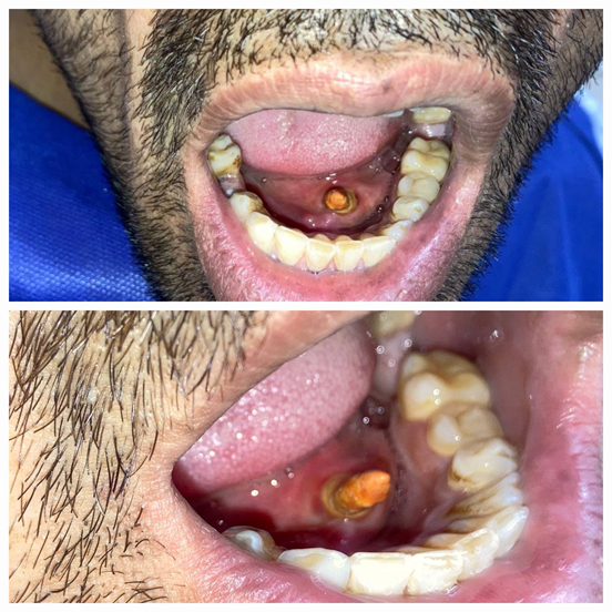
A 42-year-old male presented at the maxillofacial clinic with a persistent complaint of a sore throat on the right side that had been troubling him for the past few weeks. The patient described the discomfort as a dull, persistent ache, which worsened upon swallowing. He denied any history of fever, difficulty swallowing, or changes in voice. His past medical history was unremarkable, and he reported no significant trauma to the head or neck region. Furthermore, he had no known history of chronic conditions, such as diabetes or autoimmune diseases, nor any medications that might predispose him to salivary gland issues. Upon initial physical examination, mild swelling and tenderness were palpated in the right submandibular region, located just below the angle of the jaw. The swelling was firm to palpation and non-fluctuant, which suggested a chronic inflammatory process rather than an acute infection. Intraoral examination revealed a noticeable, firm, and irregular mass in the floor of the mouth, just beneath the tongue (Fig. 1). This finding was accompanied by mild erythema and slight swelling of the mucosa overlying the submandibular duct. There was no evidence of purulent drainage, suggesting that the duct was not acutely infected at the time of examination. Interestingly, despite having visited multiple healthcare providers for the same complaint over the course of several weeks, none had performed a thorough intra-oral examination. Previous investigations had involved imaging studies, such as a CT scan of the face and neck, which revealed a suspicious opacity in the region of the submandibular duct; however, no definitive diagnosis was made. It was only after a more focused clinical examination that a submandibular duct calculus was suspected. Given the location of the palpable mass and the patient's symptoms, the diagnosis of a salivary stone (sialolith) was considered. Submandibular duct stones are a relatively common cause of chronic, localized pain and swelling in the floor of the mouth. However, their clinical presentation can be subtle and may mimic other more common conditions, such as a sore throat, making diagnosis challenging. The presence of a stone is often confirmed with radiographic imaging, and in this case, a non-contrast intraoral radiograph, or sialography, would have been helpful in confirming the location and size of the stone, although a CT scan had already suggested the presence of an obstructing lesion. Given the clinical suspicion, the patient was counseled regarding the possible diagnosis and treatment options. Under local anesthesia, an intraoral approach was chosen for stone removal, which is generally preferred due to its minimally invasive nature. The stone, measuring approximately 1.5 cm in diameter, was successfully excised from the submandibular duct with minimal complications (Fig. 2).
The stone was grayish in color and had a firm, calcified consistency, typical of a mature sialolith. The procedure was well tolerated, and the patient was provided with post-operative care instructions, including the use of warm compresses to the affected area to reduce swelling and pain. Antibiotics and painkillers were prescribed for five days to prevent infection and manage post-operative discomfort. Salivary stimulants, such as sour candies or lemon gargles, were also recommended to aid in the recovery of normal salivary function. Postoperatively, the patient reported immediate relief from his sore throat symptoms, with a significant reduction in the swelling and tenderness in the submandibular region. The resolution of the sore throat, which had been the primary symptom causing the patient concern, was particularly striking. At the three-month follow-up visit, the patient had fully recovered, with no residual swelling or discomfort in the submandibular region. The patient was able to resume normal eating and speaking without difficulty, and no further complications were observed.
Salivary stones, or sialoliths, are calcified masses that can obstruct the ducts of the salivary glands, resulting in various clinical manifestations. This case report details the rare occurrence of a large submandibular duct calculus in a 42-year-old male who initially presented with sore throat symptoms. The patient’s clinical history, physical examination, imaging findings, and treatment approach are thoroughly outlined. This case underscores the necessity of including salivary stones in the differential diagnosis when managing patients with unusual symptoms in the head and neck region, particularly in those with atypical sore throat presentations.
Key words: Salivary Duct Calculi, Salivary Gland Calculi, Submandibular Gland
Introduction
Sialolithiasis, also known as salivary gland stones, is an organic calcified substance that forms in the paranchyma and ducts of both major and minor salivary glands; however, it usually involves a single gland. Mechanical factors that impede saliva flow, along with the physicochemical properties of gland secretions, contribute to the formation of a central core for calcification and subsequent deposition of calcium salts and phosphates. The condition is most common in adult males aged 30 to 60, with a lower prevalence in children. The precise etiology remains unclear, though potential contributing factors include inflammation, infection, anatomical irregularities in the salivary duct system, and smoking (1, 2).
Sialolithiasis predominantly affects the submandibular gland and its duct due to its complex structure, while less common occurrences are seen in the parotid gland and the sublingual or minor salivary glands. The clinical importance lies in recognizing these patterns, particularly when faced with atypical presentations that may challenge the initial diagnosis (3). It should be noted that the mucoid nature of submandibular gland secretions with high levels of calcium and phosphate makes sialolithiasis more common in this gland. Secretions from submandibular and parotid glands are nerve-dependent; accordingly, when nerve stimulation is absent, the risk of sialolith formation increases. The size of these calcified stones varies greatly (4).
Multiple stones in more than one salivary gland are scarce (about 3%). They can have various shapes, such as spherical, oval, or irregular. Symptoms can also vary, and some patients may be asymptomatic while others may experience severe pain, especially when eating. Saliva production may decrease in some patients. Swelling and inflammation around the duct entrance, oral mucosa floor, and skin near the salivary gland area may occur (5).
The severity of symptoms is influenced by the location and extent of saliva flow obstruction. Sialoliths typically range from 5 to 10 millimeters, with stones larger than 10 millimeters considered abnormally large. Stones exceeding 15 millimeters are classified as giant and are rare. Blockages can lead to chronic inflammation or acute infection. Salivary stones must be accurately identified with high precision to differentiate them from other diseases so that a specific treatment plan can be implemented for them (4-7).
While salivary stones are a relatively common condition, they can present with a wide range of symptoms, making diagnosis challenging in some cases (6). Here, we present a unique case of a large submandibular duct calculus in a middle-aged man whose initial complaint of a sore throat ultimately led to the discovery of this uncommon etiology.
Case
This case report was conducted as part of a research study to investigate the clinical and demographic characteristics of patients with facial trauma in the Emergency Department of Khatam-al-Anbia Hospital, Zahedan, Iran, in 1402 (IR.ZAUMS.REC.1402.356).
A 42-year-old male presented at the maxillofacial clinic with a persistent complaint of a sore throat on the right side that had been troubling him for the past few weeks. The patient described the discomfort as a dull, persistent ache, which worsened upon swallowing. He denied any history of fever, difficulty swallowing, or changes in voice. His past medical history was unremarkable, and he reported no significant trauma to the head or neck region. Furthermore, he had no known history of chronic conditions, such as diabetes or autoimmune diseases, nor any medications that might predispose him to salivary gland issues. Upon initial physical examination, mild swelling and tenderness were palpated in the right submandibular region, located just below the angle of the jaw. The swelling was firm to palpation and non-fluctuant, which suggested a chronic inflammatory process rather than an acute infection. Intraoral examination revealed a noticeable, firm, and irregular mass in the floor of the mouth, just beneath the tongue (Fig. 1). This finding was accompanied by mild erythema and slight swelling of the mucosa overlying the submandibular duct. There was no evidence of purulent drainage, suggesting that the duct was not acutely infected at the time of examination. Interestingly, despite having visited multiple healthcare providers for the same complaint over the course of several weeks, none had performed a thorough intra-oral examination. Previous investigations had involved imaging studies, such as a CT scan of the face and neck, which revealed a suspicious opacity in the region of the submandibular duct; however, no definitive diagnosis was made. It was only after a more focused clinical examination that a submandibular duct calculus was suspected. Given the location of the palpable mass and the patient's symptoms, the diagnosis of a salivary stone (sialolith) was considered. Submandibular duct stones are a relatively common cause of chronic, localized pain and swelling in the floor of the mouth. However, their clinical presentation can be subtle and may mimic other more common conditions, such as a sore throat, making diagnosis challenging. The presence of a stone is often confirmed with radiographic imaging, and in this case, a non-contrast intraoral radiograph, or sialography, would have been helpful in confirming the location and size of the stone, although a CT scan had already suggested the presence of an obstructing lesion. Given the clinical suspicion, the patient was counseled regarding the possible diagnosis and treatment options. Under local anesthesia, an intraoral approach was chosen for stone removal, which is generally preferred due to its minimally invasive nature. The stone, measuring approximately 1.5 cm in diameter, was successfully excised from the submandibular duct with minimal complications (Fig. 2).
The stone was grayish in color and had a firm, calcified consistency, typical of a mature sialolith. The procedure was well tolerated, and the patient was provided with post-operative care instructions, including the use of warm compresses to the affected area to reduce swelling and pain. Antibiotics and painkillers were prescribed for five days to prevent infection and manage post-operative discomfort. Salivary stimulants, such as sour candies or lemon gargles, were also recommended to aid in the recovery of normal salivary function. Postoperatively, the patient reported immediate relief from his sore throat symptoms, with a significant reduction in the swelling and tenderness in the submandibular region. The resolution of the sore throat, which had been the primary symptom causing the patient concern, was particularly striking. At the three-month follow-up visit, the patient had fully recovered, with no residual swelling or discomfort in the submandibular region. The patient was able to resume normal eating and speaking without difficulty, and no further complications were observed.
Figure. 1. Image showing the patient’s salivary stone
Figure 2. Image showing the patient after the treatment
Discussion
Salivary gland inflammation due to stones is common, especially in the submandibular gland. Determining the true prevalence of salivary stones can be challenging, as they are often asymptomatic. Factors contributing to stone formation include saliva flow issues and composition. Duct irregularities, inflammation, dehydration, and certain medications lead to flow issues, while calcium levels and a lack of crystallization inhibitors affect saliva composition. Bacterial infections can also promote stone formation by increasing saliva saturation with calcium phosphate due to pH changes. Factors, such as duct shape, calcium levels, gland position, mucus, smoking, trauma, infection, and inflammation, contribute to salivary stasis. The recurrence rate is about 20%. The stone formation mechanism is unclear but likely involves microcalcifications due to reduced secretion over time in ducts (1, 7, 8).
Salivary stones can lead to intermittent swelling, with the severity often depending on the degree of obstruction and the presence of infection. While symptoms often occur during meals, this was not the case with our patient. Chronic cases may exhibit fistulas or ulcers, which were absent in our case. Submandibular stones are generally singular, whereas sublingual stones tend to be multiple and scattered. If untreated, stones can grow larger and eventually lead to calcification. Obstruction can also trigger infections, with symptoms including pain, swelling during meals, and infection-related symptoms in the case of larger stones (1, 3, 7).
In the acute phase, treatment for sialoliths focuses on supportive care, such as pain relief, hydration, and antipyretics, along with antibiotics to address potential infections caused by salivary gland inflammation. Stimulating saliva flow and localized massage therapy are commonly employed, with treatment options ranging from minimally invasive to more radical interventions depending on the size and characteristics of the stone (5, 9).
Factors influencing treatment decisions include the duration of obstruction, tissue changes, and stone location. Non-invasive methods, such as massage and medications, are effective for stones located more distally, while deeper stones often require surgical intervention. For submandibular gland stones, lithotripsy ultrasound is a well-established treatment. Sialendoscopy and laser lithotripsy can also be used for stones of varying sizes. In the differential diagnosis of masses in the floor of the mouth, it is essential to consider conditions, such as ranula, cysts, tumors, and calcifications in the lymph nodes, ducts, and glands (1, 3, 5).
Conclusions
Salivary stones are a known cause of salivary gland dysfunction and can present with a variety of clinical manifestations, including pain, swelling, and infection. However, the presentation of a salivary stone as a sore throat is uncommon and can mimic other more common conditions, necessitating a high index of suspicion for accurate diagnosis. This case underscores the importance of considering salivary stones in the differential diagnosis of patients presenting with atypical symptoms in the head and neck region. Prompt recognition and appropriate management of salivary stones can lead to favorable outcomes and symptom resolution. Further studies and case reports are warranted to enhance our understanding of the diverse presentations and optimal management strategies for this condition.
Conflict of Interest
The authors declare no conflicts of interest.
Type of Study: Case Report |
Subject:
Oral and Maxillofacial
Received: 2024/11/27 | Accepted: 2025/06/28 | ePublished ahead of print: 2025/07/19 | Published: 2025/09/18
Received: 2024/11/27 | Accepted: 2025/06/28 | ePublished ahead of print: 2025/07/19 | Published: 2025/09/18
References
1. Pachisia S, Mandal G, Sahu S, Ghosh S. Submandibular sialolithiasis: A series of three case reports with review of literature. Clin Pract. 2019;9(1):32-7. [DOI:10.4081/cp.2019.1119]
2. Sigismund PE, Zenk J, Koch M, Schapher M, Rudes M, Iro H. Nearly 3,000 salivary stones: some clinical and epidemiologic aspects. Laryngoscope. 2015;125(8):1879-82. [DOI:10.1002/lary.25377]
3. Barrueco AS, Cornejo MVL-A, Rueda IA, Andrés SL, Galán FG, Tapia GD, et al. Sialolithiasis: Mineralogical composition, crystalline structure, calculus site, and epidemiological features. Br J Oral Maxillofac Surg. 2022;60(10):1385-90. [DOI:10.1016/j.bjoms.2022.08.005]
4. Sigismund P, Zenk J, Koch M, Schapher M, Rudes M, Iro H. Clinical and epidemiologic aspects of sialolithiasis: a retrospective study. 2015.
5. Ashindoitiang JA, Nwagbara VIC, Ugbem T, Odoks RK, Udo Solomon A, Akpan SO, et al. Huge sialolith of the submandibular gland: a case report and review of literature. J Int Med Res. 2023;51(1):03000605221148443. [DOI:10.1177/03000605221148443]
6. Pousya VS, Shetty U, Shetty P. Sialolith with Sialadenitis: A Case Study and a Proposal of a Diagnostic Algorithm. J Health Allied Sci NU. 2024;14(2):281-9. [DOI:10.1055/s-0043-1768590]
7. Basra AS, Mohod S, Shinde SB, Phaye LD, Khandelwal P. Management of a Submandibular Sialolith: A Case Report. Cureus. 2024;16(6):e61812. [DOI:10.7759/cureus.61812]
8. Rapriya A, Kumar J. A Large Size Sialolith in the Wharton's Duct: A Rare Case Report. J Res Dent Maxillofac Sci. 2024;9(2):129-31. [DOI:10.61186/jrdms.9.2.129]
9. Koch M, Mantsopoulos K, Müller S, Sievert M, Iro H. Treatment of sialolithiasis: what has changed? An update of the treatment algorithms and a review of the literature. J Clin Med. 2021;11(1):231-44. [DOI:10.3390/jcm11010231]
Send email to the article author
| Rights and permissions | |
 |
This work is licensed under a Creative Commons Attribution-NonCommercial 4.0 International License. |

Attribution-NoneCommercial CC BY-NC
