


BibTeX | RIS | EndNote | Medlars | ProCite | Reference Manager | RefWorks
Send citation to:
URL: http://jsurgery.bums.ac.ir/article-1-175-en.html


Laparoscopic treatment of a huge missed ureteropelvic junction obstruction in a 42-year-old adult: A case report study
Seyed Mehdi Saadati1![]() , Ramin Haghighi2
, Ramin Haghighi2![]() , Bagher Moradi3*
, Bagher Moradi3*![]()
1Msc of Pediatric Nursing, Imam Khomeini Hospital of Medical Sciences, Esfarayen, Iran
2Assistant professor, Faculty Member, Faculty of Medical Sciences, North Khorasan, Iran
3Assistant professor, FacultyMember, Esfarayen Faculty of Medical Sciences, Esfarayen, Iran
Received: January 15, 2018 Revised: February 14, 2019 Accepted: February 16, 2019
|
Abstract Ureteropelvic junction obstruction (UPJO) is an obstruction that occurs in renal pelvic-ureter junction. In case the UPJO diagnosis is confirmed, the treatment is surgical. In this regard, closed surgery (laparoscopic surgery) is currently recommended due to the small number of complications arising after this surgery. In the present study, we reported the laparoscopic treatment of a huge ureteropelvic junction obstruction in a 42-year-old man referred to a physician with a complaint of dull abdominal pain radiating to the back. After the computed tomography scan, the laparoscopy was performed under general anesthesia. Sever hydronephrotic kidney was dissected completely from adjacent organs and nephrectomy was performed for this case. Afterward, the ureter was ligated and divided at this level. The procedure was followed by the drainage of the cloudy urine entrapped in the kidney. Renal hilum was dissected and renovascular was secured and divided separately using multiple clips. The specimen was extracted using entrapment sac. This surgical management ended without any complication. The UPJO is a disorder of the urinary tract with an unknown etiology in elderly and middle-aged people. Early diagnosis and timely treatment can prevent some complications, such as poor functioning or nonfunctioning kidney. Nowadays, UPJO standard treatment is a laparoscopic pyeloplasty surgery to relieve obstruction and reconnect the ureter to the pelvis. To this end, a rapid and easy method of urine drainage is conducted to save the kidney. Key words: Kidney, Laparoscopy, Surgery, Ureteral obstruction |
Introduction
Ureteropelvic junction obstruction (UPJO) is an obstruction that occurs in the renal pelvic-ureter junction (1). The UPJO usually considered a congenital disorder, can block urine flow partially or completely (2). Patients usually present painful signs and it may come with flank pain. Other unusual symptoms of UPJO include fever, urinary tract infection, or hematuria (3).
Hydronephrosis is one of UPJO manifestations in which urine is blocked in the kidney. Similar to UPJO, hydronephrosis can be detected using sonography even in fetuses. However, other methods, such as intravenous pyelography (IVP) and nuclear scan can be employed for the diagnosis of this disease (4).
Regarding the therapeutic procedure of UPJO, patients affected with this disease should be subjected to surgical treatment since there is no chance of self-improvement. The standard surgical treatment of UPJO is pyeloplasty, an open surgical procedure for the removal of obstructive part and reconnection of the ureter to the renal. This surgical management can provide fast and easy urinary drainage and reduces the symptoms and risk of infection (2, 4). The results of excellent surgery and improvement of the blockage occur in 90-95% of cases (5, 6).
The disease does not have a specific genetic transmission although it may be observed in several members of a family. As a result, the main cause of blockage is not often clear. As mentioned earlier, the only way to manage this disease is surgery. Nowadays, closed surgery (i.e., laparoscopic surgery) is recommended due to the small number of complications arising after this surgery.
Cases
The patient was a 42-year-old man referred to a physician with a complaint of vague abdominal pain radiating to the back. Ultrasonography (US) and computed tomography (CT) scan were performed and a huge mass was detected on the right side of the abdomen (Figure1).
Under general anesthesia and in supine poison, a 10Fr port was inserted in umbilicus as open access. Afterward, pneoumoperitoen was established, the position was changed to flank, and two other ports, (10 Fr in supraumblicus and 5 Fr in right pararectus) were inserted in this regard. The performance of laparoscopy indicated a huge distended pelvicalyceal occupying the right half of abdomen and somewhat pelvis. Sever hydronephrotic kidney was dissected completely from adjacent organs (Figure 2).The ureter was ligated and divided at this level. The procedure was followed by the drainage of the cloudy urine entrapped in the kidney. Renal hilum was dissected and renovascular was secured and
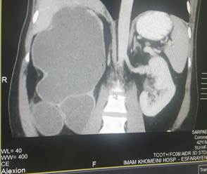
Figure1: Detection of a huge mass on the right side of the abdomen

Figure 2: Complete dissection of the kidney from the body
divided using multiple clips. The specimen was extracted using entrapment sac. The surgical management ended without any complication.
Discussion
In the present study, a 42-year-old male patient referred to a physician with a complaint of dull abdominal pain radiating to the back. The obtained results of the CT scan showed ureteropelvic junction obstruction in the patient. As prenatal screening improves, there is a decrease in the number of adult patients diagnosed with congenital UPJO (7). Most patients undergoing UPJO treatment are presented with symptoms, such as pain, infection, or urolithiasis.
It is essential to perform the diagnosis at a young age. The diagnosis of UPJO can be postponed in only a few cases. In a study conducted in 2010, a 7-year-old girl with mild flank pain and lower abdominal pain referred to the center. The clinical examination indicated painful masses around the navel with mild tenderness. Laboratory findings included kidney function tests, urinalysis, and normal culture. The renal abdominal ultrasound turned to the left and right pelvic kidney with severe hydronephrosis and normal ureter. After the necessary diagnostic procedures were performed, UPJO was diagnosed in the right ectopic kidney with normal bladder and the patient was subjected to laparoscopic pulmonary pyeloplasty (5).
The performance of laparoscopy indicated a huge distended pelvicalyceal occupying the right half of abdomen and somewhat pelvis. Sever hydronephrotic kidney was dissected completely from adjacent organs. The procedure was followed by the drainage of the cloudy urine entrapped in kidney and specimen was extracted using entrapment sac. The surgical management ended without any complication and nephrectomy was performed for this case. It should be mentioned that there are several reports in which the patients were children; however, UPJO in middle-aged patients is rare. For this reason, this study was conducted to report UPJO in a 42-year-old patient. Physicians can consider UPJO as a factor in abdominal pain and mass in emergency referrals. It should be noted that the obstruction of the ureter to the pelvis is one of the issues that should be distinguished from other pelvic masses.
Saghafi et al. (2015) reported the rare case of a pelvic mass. The patient was a single 18-year-old woman with an irregular complaint of menstruation for one year with an ultrasound diagnosis of ovarian cyst. The primary ultrasound showed the cyst was about 2×12 cm in the right lumbar spine. After 3months of taking contraceptive pills, another cyst with the dimensions of 68×95×98 mm with a possible source of right adnexa was reported in the ultrasound. Given the abdominal pain and the large size of the cyst, the patient was a candidate for UPJO laparotomy. The origin of the cyst was the kidney pelvis. The right ectopic kidney containing the pyeloplasty was treated using Anderson Hayes method as well as the double-J-catheter (8).
In another case study reported by George et al. (2009), a 28-year-old woman referred with abdominal pain complaints and a large cyst on the left side of the lower pelvis, which imitated a multicystic ovarian tumor in ultrasound. The laparoscopic assessment showed that the uterus and ovaries were normal and inflammation was observed in the retroperitoneal region of the left side that was detected after opening the peritoneal space of the large hydatid cyst (9). Therefore, all different types of the pelvic mass diagnosis, especially with the urinary and digestive tract, should be taken into account and the necessary diagnostic measures should be taken before the surgery.
Conclusions
The UPJO is an unusual disorder of the urinary tract with an unknown etiology in elderly and middle-aged people. Early diagnosis and timely treatment can prevent the development of such disease. Nowadays, UPJO standard treatment is
a laparoscopic pyeloplasty surgery to relieve obstruction and reconnect the ureter to the pelvis. To this end, a rapid and easy method of urine drainage is conducted to save the kidney.
Conflict of Interest
Authors declare no conflicts of interest.
References
1. Park JM, Bloom DA. The pathophysiology of ureteropelvic junction obstruction. Urol Clin North Am. 1998; 25:161-9.
2. Gillenwater JY. Adult and pediatric urology. Philadelphia: Lippincott Williams & Wilkins; 2002.
4. Liang CC, Cheng PJ, Lin CJ, Chen HW, Chao AS, Chang SD. Outcome of prenatally diagnosed fetal hydronephrosis. J Reprod Med. 2002; 47(1):27-32. PMID: 11838306
5. Baek M, Park K, Choi H. Long-termoutcomes of dismembered pyeloplasty for midline-crossing giant hydronephrosis caused by ureteropelvic junction obstruction in children. Urology. 2010; 76(6):1463-7. PMID: 20800889 DOI: 10.1016/j.urology.2010.05.040
6. Sutherland RW, Chung SK, Roth DR, Gonzales ET. Pediatric pyeloplasty: outcome analysisbased on patient age and surgical technique. Urology. 1997; 50(6):963-6. PMID: 9426731 DOI: 10.1016/S0090-4295(97)00397-X
7. Capello SA, Kogan BA, GIorgi LJ JR, Kaufman RP JR. Prenatal ultrasound has led to earlier detection and repair of ureteropelvic junction obstruction. J Urol. 2005; 174(4 Pt 1):1425-8. PMID: 16145455 DOI: 10.1097/01.ju.0000173130.86238.39
8. Saghafi N, Porali L, Jafarian F. A rare case of a pelvic mass protest. Iran J Obstet Gynecol Inferctil. 2015; 18(143):17-21. DOI: 10.22038/IJOGI.2015.4337
9. Görgen H, Api M, Çetin A. Primary adnexial hydatid cyst mimicking ovarian tumor. J Turk Ger Gynecol Assoc. 2009; 10(4):232-4. PMID: 24591878
Received: 2019/01/15 | Accepted: 2019/02/16 | ePublished ahead of print: 2019/07/6 | Published: 2019/09/22 | ePublished: 2019/09/22
| Rights and permissions | |
 |
This work is licensed under a Creative Commons Attribution-NonCommercial 4.0 International License. |

Attribution-NoneCommercial CC BY-NC
